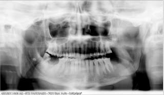
 1st opg showing osteolytic area in region of 37.not very frank radiolucency.hard to detact radiologically without clinical information because of edentulous area.clinically patient was having a small ulceroproliferative growth in the same region.incisonal biopsy suggested moderately differentiated epithelial squamous cell carcinoma.patient was refered to higher centers for mangment of malignancy.
 opg of same patient after 6 month showing displaced pathological fracture at angle of mandible.showing progressive nature of malignant process.opg was taken at aiims.
final opg u can see the severe destruction of cortex in form of diffuce radiolucency with ill defined borders.tooth also showing floating tooth appearance.patient again reported for biopsy slide for revaluation before the treatment.mri was also adviced for this patient .
No comments:
Post a Comment